

Biomedical Spectroscopy and Imaging (BSI)
The sophisticated high-level bioimaging instrument is a necessary implement for the pursuit of excellence in biomedical research. Which provides researchers to carry out qualitative and quantitative investigation of cells and molecularly-targeted in real, non-destructive, and complete in vivo. Furthermore, the image shows the physiological and pathological phenomena of cells or molecules directly, and convert the complex processes such as gene expression and biosignal transmission into images. Also, with the help of specific molecular probes, it is possible to achieve the goal of early diagnosis by discover disease symptoms under the molecular level. Therefore, it is necessary to possess a variety of biomedical optical imaging instruments with a variety of specific and special features for pursuing excellent biomedical research.
Raman Spectroscopy:

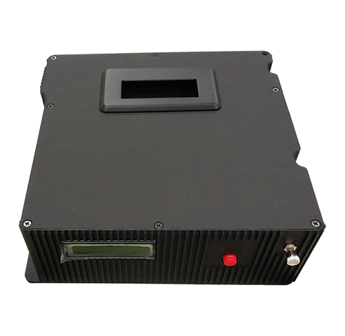
TR500 Raman Spectral


TS-AU3 SERS chip

The notable improvements in both sensitivity and imaging speed have paved the way to apply the powerful analytical capability of Raman spectroscopy for biomedical research. Raman imaging has now become a new imaging modality that can provide molecular-level information in biological systems inaccessible by conventional optical techniques.
It is used as a non-invasive and nondestructive diagnostic tool to analyse molecular changes in cells, tissues or biofluids. Changes that are either the cause or the effect of pathological diseases. Raman spectroscopy can provide a wealth of biochemical information rapidly to improve understanding and help in the prediction of disease in research and clinical settings. Compared to other molecular and imaging diagnostics techniques, Raman spectroscopy is noninvasive, fast, low-cost and can achieve high chemical specificity in biological samples.
From the trend of Raman spectroscopy development, Surface-Enhanced Raman Scattering (SERS) had been widely used in life science research. Surface-Enhanced Raman Scattering (SERS), the combination of geometry and materials gives rise to a massive enhancement of the electromagnetic field which can be matched to the laser wavelength. This amplifies the incident laser power like a radio antenna, and also acts as a transmitter to enhance the Raman-shifted signal. Enhancements as high as 10-14 have been recorded from dye molecules on silver particles, but enhancements of several orders of magnitude are typical. These gold (or silver) particles can be ‘bare’ to enhance the Raman signal of a molecule within a few nanometers of the surface of the particle.
Hyperspectral Imaging:

The goal of hyperspectral imaging is to obtain the spectrum for each pixel in the image of a scene, with the purpose of finding objects, identifying materials, or detecting processes.
Whereas the human eye sees the colour of visible light is mostly three bands (long wavelengths – perceived as red, medium wavelengths – perceived as green, and short wavelengths – perceived as blue), spectral imaging divides the spectrum into many more bands. This technique of dividing images into bands can be extended beyond the visible. In hyperspectral imaging, the recorded spectra have fine wavelength resolution and cover a wide range of wavelengths (including: UV light ~ visible light ~ infrared light bands, and even more). Hyperspectral imaging measures continuous spectral bands.
The primary advantage to hyperspectral imaging is that, because an entire spectrum is acquired at each point, the operator needs no prior knowledge of the sample, and postprocessing allows all available information from the dataset to be mined. Hyperspectral imaging can also take advantage of the spatial relationships among the different spectra in a neighbourhood, allowing more elaborate spectral-spatial models for more accurate segmentation and classification of the image.
